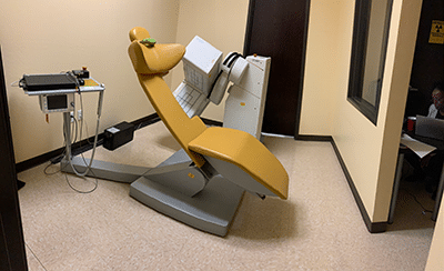
Nuclear Cardiology
Nuclear imaging evaluates how organs function, unlike other imaging methods that assess how organs appear. Small amounts of a radioactive solution(s) are introduced into the body. A special camera detects the solution in different parts of the body and a computer generates a series of images of the areas of interest.
Nuclear cardiac imaging can help determine if there is adequate blood flow to the heart muscle during stress versus rest. It can also evaluate heart function and the presence of prior heart attacks.
Cardiac nuclear images (also known as SPECT imaging – Single Photon Emission Computed Tomography) help to identify coronary heart disease, the severity of prior heart attacks, and the risk of future heart attacks. These highly accurate evaluations of cardiac function and amount of heart muscle at risk enable cardiologists to better prescribe medications and select further testing like a coronary angiogram, the need for angioplasty and bypass surgery, or devices to optimize treatment outcomes.
Nuclear Cardiology Services at San Tan Cardiovascular Center
At our state-of-the-art facilities, we perform approximately 1,000 nuclear cardiac imaging studies annually. Our nuclear cardiology laboratory performs a variety of non-invasive cardiovascular imaging studies:
MUGA Scan
- Radionuclide cineangiograms (RNCA/MUGA studies) to evaluate heart function
Nuclear Stress Test
- Myocardial perfusion imaging for the detection and management of coronary artery disease
- Viability studies to assess for the extent of myocardial infarction
Stress modalities routinely performed include:
- Exercise treadmill testing
- Vasodilator pharmacologic stress
- Dobutamine stress testing.

Nuclear Medicine at our Mesa, AZ location
Our gamma cameras can acquire a complete study in half the time compared to conventional imaging protocols and we can utilize a reduced amount of the nuclear tracer. Our state-of-the-art facility is accredited by the Inter-societal Accreditation Commission (IAC/Nuclear). Our laboratory provides the highest diagnostic accuracy, while optimizing patient satisfaction, comfort and care.